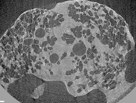
PHASE CONTRAST IMAGING
In Phase-Contrast X-ray Imaging (PCI), the change in the phase of a X-ray beam that passes through an object is recorded by specific detectors, creating projection images of the object. In respect to usual X-rays techniques, which rely on the measurement of the attenuation of X-rays interacting with matter, PCI is useful when imaging soft tissues that do not possess large variations in X-ray absorption properties. Phase Contrast Imaging can also be combined with tomographic techniques to obtain 3D reconstruction.
Applications of PCI include for example cartilage bone imaging, angiography, lung imaging, mammography.
See also: S.-A. Zhou and A. Brahme, “Development of phase-contrast X-ray imaging techniques and potential medical applications”, https://doi.org/10.1016/j.ejmp.2008.05.006
Phase Contrast Imaging is available at the Phase Contrast Imaging Flagship Node Trieste.
Click here for Use Cases from our Nodes.
Propagation based phase contrast CT
Propagation-based phase-contrast imaging uses the free-space propagation of a coherent X-ray beam to create contrast. After passing through the sample the wavefront is distorted as a consequence of the phase-shift imposed by the sample. The propagation of the distorted X-ray wavefront gives rise to Fresnel diffraction fringes in the image which enhance edges and interfaces present in the sample. This edge enhancement is one of the key features of phase contrast CT images, and it is especially useful for weakly-attenuating samples, which are otherwise not seen in the image.

Analyzer Based CT
In the analyser-based imaging approach (ABI) the incident X-ray beam is refracted by a crystal, typically with a bandwidth in the micro-radiant range, placed between the sample and the detector. The crystal behaves as an angular filter selectively accepting only the X-rays that have traversed the sample following Bragg diffraction.
Typically the setup to perform ABI can be found in a synchrotron facility.
In preclinical small animal imaging, ABI has been used e.g. to investigate cartilage abnormalities of degenerative joint disease.