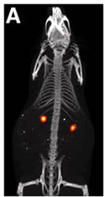
Magnetic Particle Imaging (MPI)*
Magnetic particle imaging is a tomographic method based on detection of nonlinear response of superparamagnetic tracers (usually superparamagnetic Iron Oxide, SPIO) to alternating magnetic fields.
The method detects the tracer only (i.e., it is a hot spot imaging). Therefore MPI images have no background signal, but require colocalization with an anatomical imaging method, such as MRI or CT. At moderate spatial resolution, MPI provides high sensitivity and superb temporal resolution.
Current main applications are in the preclinical field and include biodistribution of nanoparticles, cell tracking, detection of nanoparticles deployed for thermoablation.

Magnetic Particle Imaging is provided by the Centre for Advanced Preclinical Imaging Prague (CAPI)