SPAIN
Barcelona Super-resolution Light and Nanoscopy Node
The Barcelona Super-resolution Light and Nanoscopy Node (SLN@BCN) is a multi-site bioimaging Node that brings together two core facilities in Barcelona, Spain. The Node offers a collection of state-of-the-art super-resolution imaging techniques, along with a wide range of complementary and advanced imaging technologies. SLN@BCN has the expertise and infrastructure for experimental design, sample preparation, acquisition, as well as the processing and analysis of the complex datasets generated by the acquired images. The Node also has the capacity to provide customized bioimage analysis based on project requirements. Each unit within the SLN@BCN Node is actively involved in organizing training courses focused on both theoretical and practical aspects of basic and advanced microscopy techniques and bioimage analysis.
Specialties and expertise of the Node
At SLN@BCN, we perform research and development at the cutting-edge of several microscopy and super-resolution imaging techniques to continually improve the specifications of our microscopes, aiming to provide unique features that adapt to project needs. We collaborate with industry, hospitals and research centres. Our research covers a wide range of applications, including the visualization of single molecules, subcellular components, and cells. The SLN@BCN Node provides the required infrastructure for storing generated data and tailored data analysis pipelines, as well as secure data transfers between organizations.
Offered Technologies
| Technologies | Euro-BioImaging |
| Deconvolution widefield microscopy (DWM) | ✓ |
| Single Molecule localisation microscopy (SMLM) | ✓ |
| Reversible saturable optical fluorescence transitions (RESOLFT) | ✓ |
| Laser scanning confocal microscopy (LSCM / CLSM) | ✓ |
| Spinning disc confocal microscopy systems (SDCM) | ✓ |
| Deconvolution widefield microscopy (DWM) | ✓ |
| Total internal reflection fluorescence microscopy (TIRF) | ✓ |
| Two-photon microscopy (2P) | ✓ |
| Second/Third Harmonic Generation | ✓ |
| Image Scanning Microscopy (ISM) | ✓ |
| Fluorescence-lifetime imaging microscopy (FLIM) | ✓ |
| Fluorescence Resonance Energy Transfer (FRET) | ✓ |
| Fluorescence Recovery After Photobleaching (FRAP) | ✓ |
| Raman spectroscopy (RS) | ✓ |
| High throughput microscopy/high content screening (HTM/HCS) | ✓ |
| Image Analysis-bio | ✓ |
Additional Services offered by the Node
- Instruments
- Technical assistance to run instrument
- Methodological setup (e.g. design of study protocol and standard operation procedures)
- Training in infrastructure use
- Probe preparation
- Wet lab space
- Server Space
- Data processing and analysis
- Training workstations
- Training seminar room
- Logistics support for housing facilities
- Cell culture facilities - Safety level 1
- Cell culture facilities - Safety level 2
- Mechanical and electronic workshop
- Micro- and macro-fabricated parts through rapid prototyping
Instrument highlights
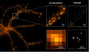
STORM: The Nikon N-STORM system is equipped with 4 laser lines (405, 488, 561, 647nm), a cage stage incubator for live-cell experiments (facilitating PALM experiments) and an EMCCD. The system includes an additional CMOS (Hamamatsu Orca Flash V3), NIS-Elements AR v5 software upgrade and enhanced performances of the current system. We can perform N-STORM, dSTORM, PALM, and DNA PAINT. The system is also equipped with environmental control for temperature, humidity and CO2 for live-cell imaging.
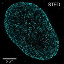
STED: The Leica TCS SP8 FALCON STED 3X is based on the TCS SP8 confocal microscope equipped with temperature control. The STED 3X module allows 3D super-resolution capabilities in 3 optical bands due to its 3 depletion lasers (592 & 660 nm CW, and 775 nm pulsed laser). The FALCON (FAst Lifetime CONtrast) is a fluorescence lifetime imaging microscopy (FLIM) platform fully integrated in the confocal microscope that can deliver video-rate FLIM with pixel-by-pixel quantification. The single molecule detector (SMD HyD) provides a high detection efficiency. Resonant scanners provide fast scanning capabilities up to video-rate at 512×512 pixels. The system includes a white light laser source as well as a 405 nm semiconductor laser. Live-cell imaging is possible due to controlled environmental conditions. We also provide STED image processing with the Huygens software.
STED: The Abberior STED microscope is based on the INFINITY platform, which is built around an Olympus IX83 microscope. The system is equipped with four pulsed lasers (485, 518, 561, 640nm) as well as a CW 405nm laser. Moreover, the STED module has two STED depletion lasers (595, 775nm). The detection units are based on avalanche photodiodes (APDs) and we have the MATRIX detector upgrade, which consists on a hexagonal arrangement of avalanche photodiodes (APDs) enabling a higher signal-to-background. Moreover, we have an additional spectral detection unit including ultra-high sensitivity FLIM optimized APD.
Airyscan: The LSM980 airyscan 2 and definite focus 2 microscope is equipped with temperature, humidity and CO2 control for live-cell imaging. The system is equipped with several objectives including silicon, water, glycerol and oil to accommodate various sample refractive indices and multiple laser lines (405, 488, 561 and 639 nm). The airyscan’s multiplex mode allows to choose from or find a compromise between super-resolution, fast or better contrast imaging.
Contact details
Dr. Pablo Loza-Alvarez
ICFO- The Institute of Photonic Sciences
Super Resolution Light Microscopy and Nanoscopy (SLN) Lab
Mediterranean Technology Park
Av. Carl Friedrich Gauss, 3
08860 Castelldefels (Barcelona), Spain
Tel: +34 93 55 34 075
e-mail: PabloLoza@icfo.eu
http://www.icfo.es
http://sln.icfo.eu
Dr. Nadia Halidi
Advanced Light Microscopy Unit (ALMU)
Centre for Genomic Regulation (CRG)
Doctor Aiguader, 88,
08003 Barcelona, Spain
Tel: +34 93 316 03 72
e-mail: nadia.halidi@crg.eu
www.crg.eu
www.crg.eu/en/programmes-groups/advanced-light-microscopy-unit