European Molecular Biology Laboratory (EMBL)
Euro-BioImaging EMBL-Node
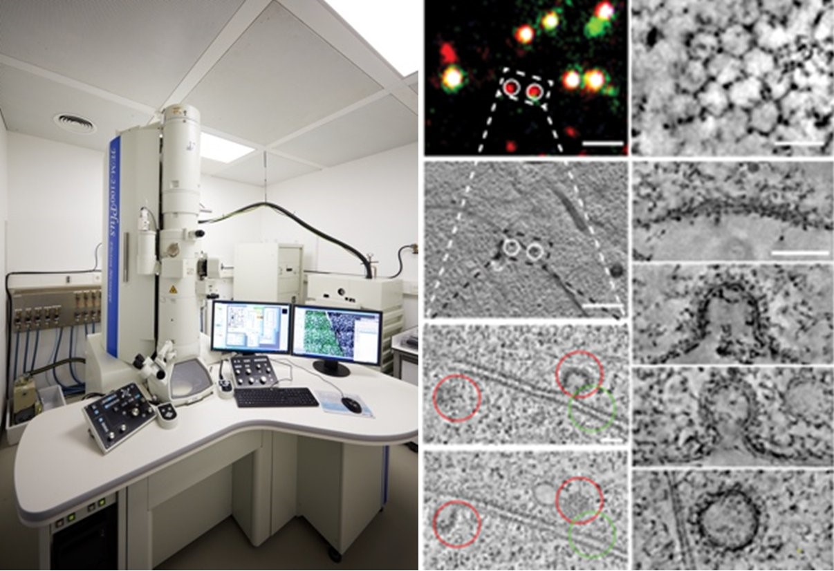
The Euro-BioImaging EMBL-Node offers a collection of state-of-the-art microscopy equipment and image processing tools. The facility was set up as a cooperation between EMBL and industry to improve communication between users and producers of high-end microscopy technology. This Node supports in-house scientists and visitors in using microscopy methods for their research and regularly organizes in-house and international courses to teach basic and advanced microscopy methods. The services provided include project planning, sample preparation, microscope selection and use, image processing and visualization. The Euro-BioImaging EMBL-Node supports advanced microscopy techniques such as FRAP, FRET, FCS, high-throughput microscopy, laser nanosurgery, CLEM, mesoscopy and super-resolution microscopy.
Specialties and expertise of the Node
The Euro-BioImaging EMBL-Node offers its expertise in the development of user defined comprehensive workflows in automated high throughput microscopy, including automated sample preparation, image acquisition, image analysis and data mining. This technology is most typically applied by users to large-scale projects, e.g. genome-scale siRNA screening. Based on its experience in high throughput microscopy and image analysis the Euro-BioImaging EMBL-Node is able to offer approaches, that allow scanning of the sample for objects of interest by fast image acquisition and online image analysis followed by more detailed automated imaging such as multi-color 3D time-lapse microscopy, FRAP or FC(C)S.

Offered Technologies:
| Technologies |
| Deconvolution widefield microscopy (DWM) |
| Laser scanning confocal microscopy (LSCM/CLSM) |
| Spinning disk confocal microscopy (SDCM) |
| Total internal reflection fluorescence microscopy (TIRF) |
| Two-photon microscopy (2P) |
| Image Scanning microscopy (ISM) |
| cryoFM * |
| Single Molecula localisation microscopy (SMLM) |
| Stimulated emission depletion microscopy (STED) |
| Reversible optical fluorescence transitions (RESOLFT) |
| Minimal Photon Fluxes Microscopy (MINFLUX)* |
| Light-sheet mesoscopic imaging (SPIM or dSLSM) |
| Optical projection tomography (OPT) |
| High throughput microscopy/high content screening (HTM/HCS) |
| Intravital Microscopy (IVM) |
| Fluorescence (cross)-correlation spectroscopy (FCS/FCCS) |
| Fluorescence Resonance Energy Transfer (FRET) |
| Fluorescence Recovery After Photobleaching (FRAP) |
| Fluorescence Lifetime Imaging Microscopy (FLIM) |
| Microdissection * |
| Imaging at Biosafety Level >1 |
| TEM of chemical fixed samples (TEM) |
| TEM of cryo-immobilized samples (TEM cryo samples)* |
| Large scale EM |
| EM tomography (ET) |
| serial section TEM |
| Serial Blockface SEM |
| Focussed Ion beam SEM (FIB-SEM) |
| Immuno-gold EM on thawed cryo-sections (Tokuyasu-EM) |
| Immuno-gold EM on resin sections (resin-EM) |
| pre-embed CLEM |
| pre-embeded CLEM |
| on-section CLEM |
| Scanning Electron Microscopy (SEM) |
| live-cell CLEM |

Additional services offered by the Node
- Methodological setup (e.g. design of study protocol and standard operation procedures)
- Technical assistance to run instrument
- Training in infrastructure use
- Probe preparation
- Animal preparation
- Animal facilities
- Wet lab space
- Server space
- Data processing and analysis
- Training workstations
- Training seminar rooms
- Housing facilities
- Biological material storage and processing
- Training in techniques for optical clearing of biological samples
Please note that not all technologies & services are available at all node sites.

Contact details
Dr. Rainer Pepperkok
Head of Advanced Light Microscopy Facility
+4962213878332