MEG – key imaging technology for understanding neuronal processing directly in a non-invasive fashion
Published: 2021-09-28
Tell us who you are, where your facility is based, and what your role is.
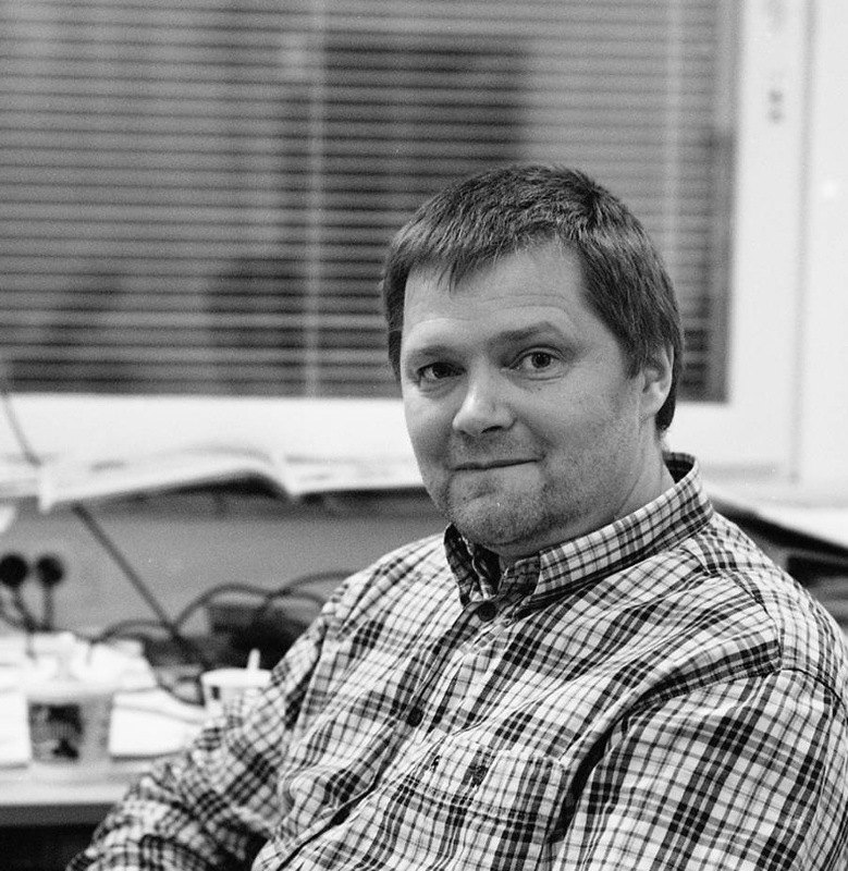
I am Veikko Jousmäki, physicist, senior scientist, Head of Aalto NeuroImaging open-access research infrastructure for human functional neuroimaging at Aalto University, Espoo, Finland.
My role is to maintain and develop Aalto NeuroImaging facility together with my team. Aalto NeuroImaging comprises of four units offering magnetoencephalography (MEG), functional magnetic resonance imaging (fMRI), navigated transcranial magnetic stimulation (nTMS), and behavioural measures. We focus on neuroimaging studies in healthy subjects. We provide hands-on support in experimental designs, data acquisition, and data analysis.
My main interest is to design new stimulators and monitoring devices for functional neuroimaging to investigate cortical processing.


We are today talking about Magnetoencephalography (MEG). Please provide a short summary of this type of imaging and list some applications:
Summary: Magnetoencephalography, MEG, measures minute magnetic fields of the order of pT produced by neuronal currents of the brain. The same neuronal currents give rise to electroencephalographic (EEG) signals—both technologies share ms-level temporal accuracy and they both provide direct measures of neuronal currents. MEG is optimal for detecting neuronal currents at the fissural cortex of the brain offering a direct, non-invasive measures, e.g., of the primary sensory signals and epileptic discharges. Source modelling tools are typically used in MEG to estimate the location of the neuronal activity and its strength. The same tools can also use the EEG data and in clinical use combined MEG and EEG recordings are used especially in epilepsy diagnosis.
MEG technology involves either superconducting sensors or modern optical sensors to measure magnetic fields and magnetically shielded room to attenuate ambient magnetic noise. MEG started with a single-channel superconducting MEG-system in the 70’s and the first whole-head MEG systems were commercially available in the 90’s. Nowadays, there are more than 200 MEG systems in active use globally and more than half of them are in clinical use. Optical MEG systems are developing rapidly at the moment and the first whole-head optical MEG systems will enter the market soon.
Applications: MEG can be used in a similar fashion as EEG is being used. MEG can be utilized to study unaveraged MEG data, i.e., cortical oscillations, resting state networks, and epileptic discharges. It can also be utilized to investigate evoked responses by using external stimuli in various sensory modalities. MEG has two clinical applications, i.e., localization of epileptic foci and functional landmarks in presurgical evaluations.
MEG is also widely used in basic research to investigate, e.g., sensory processing, language processing, visual and motor systems. In addition, MEG is used to find possible biomarkers for neurodegenerative diseases.
Tell us a bit more about a specific project that was done in your facility using this technology? What scientific questions were you addressing?
Professor Marta Molinas from Norwegian University of Science and Technology contacted Aalto NeuroImaging to investigate the possibilities to find neuronal correlates to their experimental design consisting of red and green visual cues. The main motivation is to localize and quantify the neuronal source on the basis of MEG recordings and then proceed to a portable EEG system. Norway does not have MEG available and thus open-access MEG facility in Nordic countries was their first choice. In addition, we just happened to upgrade our visual stimulator system enabling ms-precision visual stimulation in MEG.
Aalto NeuroImaging has contributed to a unique method, corticokinematic coherence (CKC; for a review see Bourguignon et al., 2019) and MEG-compatible proprioceptive stimulator (Piitulainen et al., 2015), which can be used to track down sensorimotor processing in detail. CKC method is now listed as one of the methods recommended by the clinical guidelines of the American Clinical MEG Society (De Tiège et al., 2020)
References:
Piitulainen et al., 2015 https://pubmed.ncbi.nlm.nih.gov/25770989/ DOI 10.1016/j.neuroimage.2015.03.006
De Tiège et al., 2020 https://pubmed.ncbi.nlm.nih.gov/33165229/ DOI 10.1097/WNP.0000000000000481
Bourguignon et al. 2019 https://pubmed.ncbi.nlm.nih.gov/31513941/ DOI 10.1016/j.neuroimage.2019.116177
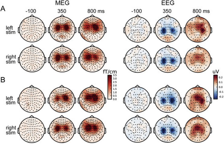
Another recent study involved comparing detection of the cortical ~20-Hz rhythm, used to evaluate the functional state of the sensorimotor cortex, by both magnetoencephalography (MEG) and electroencephalography (EEG), in healthy subjects. We expect to find the method valuable during rehabilitation, e.g., in stroke patients.

Topographic maps showing group averaged (n = 24) field strengths of the ~20-Hz rhythm modulation to (A) tactile and (B) proprioceptive stimulation both in MEG and EEG (magnetic field vs. electric scalp potential). Note that MEG topoplots shows vector sums of gradiometers (positive value) in each location.
https://doi.org/10.1016/j.neuroimage.2020.116804.
Read the full article:
Mia Illman, Kristina Laaksonen, Mia Liljeström, Veikko Jousmäki, Harri Piitulainen, Nina Forss,
Comparing MEG and EEG in detecting the ~20-Hz rhythm modulation to tactile and proprioceptive stimulation, NeuroImage, Volume 215, 2020, 116804, ISSN 1053-8119, https://doi.org/10.1016/j.neuroimage.2020.116804.
(https://www.sciencedirect.com/science/article/pii/S1053811920302913)

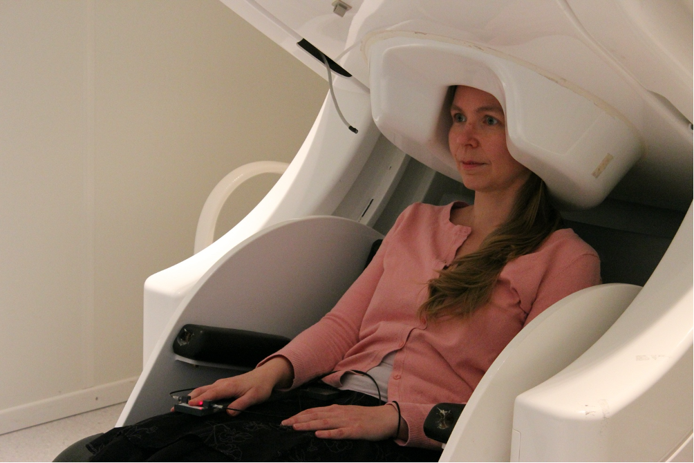
A typical MEG measurement setup used in active finger lifting motor task. A helmet-shaped MEG sensor array with 306 channels measures MEG signals while finger lifts are monitored with a fiber-optic sensor. MEG measurements can be combined, e.g., with simultaneous electroencephalographic (EEG), electro-oculograpic (EOG), electrocardiographic (EKG), electromyographic (EMG), and accelerometric signals. In addition, eye tracking, motor responses, and utterances can be recorded synchrony with MEG.
Why is this technology best suited to address that question?
MEG and EEG share the same origin of signals. Although MEG technology is expensive it has several advantages over EEG when it comes to separating cortical sources using source modelling tools. Once the estimation of the neuronal activity is carefully done on the basis of MEG the results can be easily translated to improve the portable EEG device.
What other services do you provide in your facility that would be useful in combination with this type of imaging?
The main workhorses, MEG and fMRI devices, at Aalto NeuroImaging are provided by commercial vendors. Our speciality is in stimulation and monitoring—we have a variety of commercial and in-house stimulators and monitoring devices that are compatible with modern functional neuroimaging technologies. Our users can often use the same stimulators and paradigms in multiple neuroimaging modalities to have a better view on their neuroscientific question.
Why should scientists choose your facility for using this technology?
Our users will definitely benefit from the 30-years long tradition and experience in MEG research at our facility. We will help our users to learn about the possibilities and caveats and improve their MEG experiments. As a research infrastructure, we offer a unique open-access MEG facility with fixed hourly rates, hands-on support, and dedicated staff. And finally, we are currently in the process of upgrading our MEG system. As of February 2022, our facility will proudly offer open-access to top-of-the-line MEG via Euro-BioImaging.
How to apply:
All scientists, regardless of their affiliation, area of expertise or field of activity can benefit from Euro-BioImaging’s pan-European open access services. Potential users of these new technologies are encouraged to submit project proposals via our website. To do so, you can LogIn to access our application platform, choose the technology you want to use and the facility you wish to visit, then submit your proposal. All applications will be processed by the Euro-BioImaging Hub. As usual, users will benefit from advice and guidance by technical experts working at the Nodes, training opportunities, and data management services.
For more information: info@eurobioimaging.eu